Awesome How To Identify X Ray Report

While the images are taken you will need to stay very still and may need to hold your breath for a second or two.
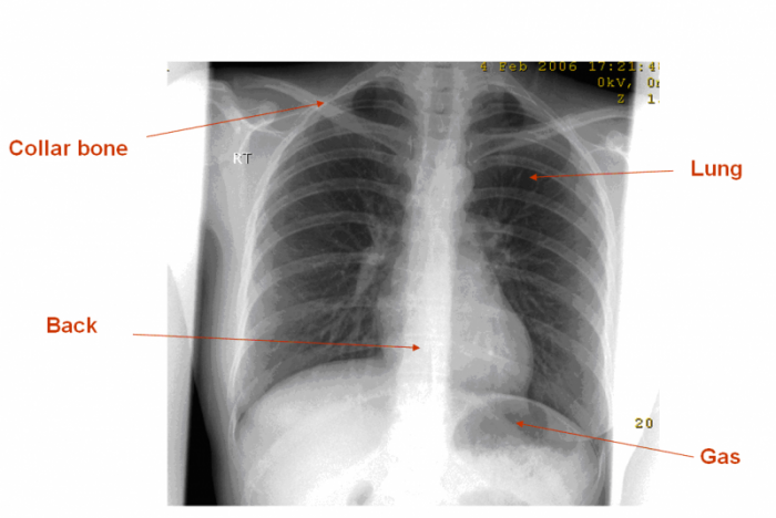
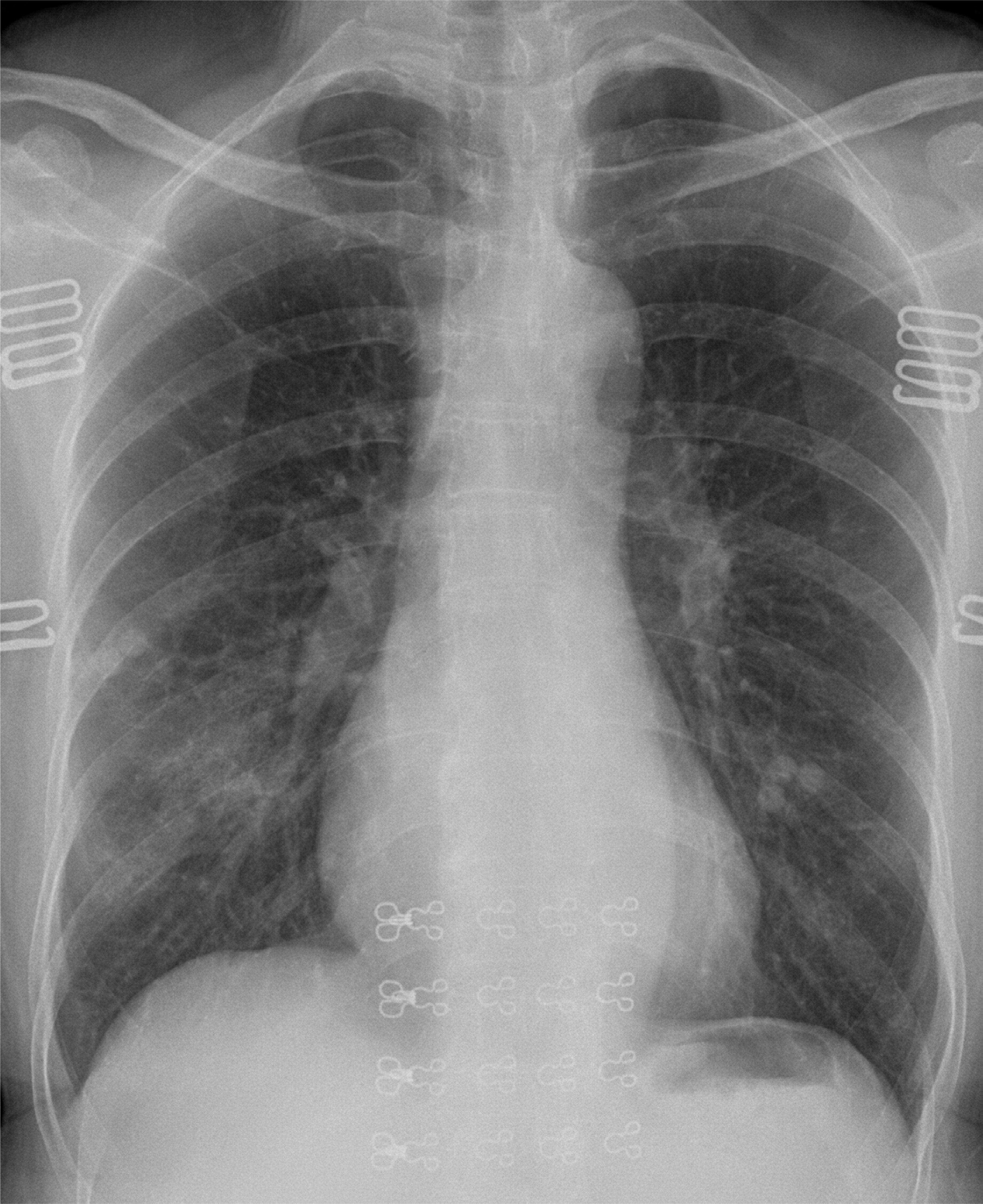
How to identify x ray report. Some physicians order an MRI before obtaining regular x-rays it is good practice to first get a regular x-rays as they are good for detecting fractures arthritis or abnormal bones in the shoulder. X-rays travelling through lung tissue cast less of a shadow than those passing through the soft tissues of the mediastinum or abdomen. In the case of pneumonia air within the alveoli is replaced by inflammatory exudate and pus.
An x-ray tech will put you in position and direct the x-ray beam to the correct part of your body. Types of x-rays white x-rays and characteristic x-rays. The left lung has three zones but only two lobes.
The anode is a water-cooled block of Cu containing desired target metal. Normal anatomy and variants. Interstitial opacities including cardiogenic and non-cardiogenic pulmonary edema and the 3 types of interstitial patterns r.
When an outer shell electron moves to fill the space created in the inner. Intensity I is either reported as peak height intensity that intensity above background or as integrated intensity the area under the peak. Look for an effusion There are two fat pads in the knee the suprapatellar fat pad the prefemoral fat Read More Knee X-rays.
An X-ray diffraction pattern is a plot of the intensity of X-rays scattered at different angles by a sample The detector moves in a circle around the sample The detector position is recorded as the angle 2theta 2θ The detector records the number of X-rays observed at each angle 2 θ The X-ray. Name and date of birth of the patient Side of extremitybody Date of x-ray Two views help to fully describe the fracture in both planes. X-ray images are grayscale.
Characteristic x-rays are caused by the ejection of an electron from an inner shell of an atom hit by the incident x-ray. The example of shoulder plain x-ray. There are OA changes seen at the first CMC joint with subchondral sclerosis and joint space narrowing.


:watermark(/images/watermark_only.png,0,0,0):watermark(/images/logo_url.png,-10,-10,0):format(jpeg)/images/anatomy_term/mediastinum-1/gee1mDStA973MGdqlBMxQ_RackMultipart20180207-1462-11chyha.png)


